The Orthopedic Clinic receives many cases, including those related to accidents such as fractures, ligament tears, muscle tears, and tendonitis. There are also cases related to the joints, some of which are chronic and some acute. Each case is dealt with according to the recommendations of the mother school for fractures, bones, and joints. Cases are dealt with academically, starting with receiving the patient, giving him his full rights, taking the medical history, and then clinical examination, x-rays, and the necessary examination for each case according to the protocol followed in all medical complexes affiliated with the Kingdom of Saudi Arabia.
We will start talking about the most common cases in the orthopedic clinic at Al Farabi Medical Complex.
Many types of bone fractures
Ligament tears are received in the orthopedic clinic, and most cases are closed fractures.
Closed fractures (also known as simple fractures) are characterized by the absence of damage to the skin around the broken bone.
Green stick fractures (most closed fractures in children are called…)
Its symptoms and signs:
- A feeling of pain or discomfort with any pressure—no matter how slight or gentle—on or around the injured area.
- Swelling of the injured area.
- The broken limb (if the fracture is in a limb) may be unable to bear any weight placed on it, and the limb may appear deformed.
- The soft tissue around the broken bone may be damaged.
How to diagnose it:-
A fracture is usually diagnosed by examining the patient and taking x-rays of the area, such as a CT scan or magnetic resonance imaging (MRI).
Treatment of fractures:-
It depends on the type and location of the fracture, as well as the patient’s age and medical history. It’s worth noting that most fractures, even minor ones, are immobilized by wrapping them in a cast, splint, or other device, depending on the patient’s needs. However, if there is severe swelling in the fracture area, the doctor may wait until the swelling subsides before applying a cast.
Compression fractures are treated solely by resting and refraining from the activity that caused them, in addition to using anti-inflammatory medications such as Voltaren, and using ice packs.
General rules for treating fractures
The injured person should remain motionless until the broken bone is immobilized.
General advice
Regular exercise is essential, as it helps prevent fractures.
Adequate amounts of calcium should be included in the diet of all ages to help prevent fractures.
Splint care
Fractures may be treated with splints to immobilize them, facilitating proper bone healing and reducing pain associated with movement. Therefore, proper care must be taken with this splint to minimize complications and prevent infection, by following the following:
- The fractured area is often swollen, and the patient may initially feel tight. To reduce swelling, elevate the splint on pillows and raise it above heart level for 24 to 48 hours.
- Ice the swollen area by placing an ice pack or a clean cloth filled with ice. It is preferable to apply the compresses for 20 minutes every two hours, avoiding applying ice directly to the skin.
- Take pain medication for at least 48 hours, such as acetaminophen or ibuprofen, to relieve pain.
- Keep the splint dry while bathing and avoiding water from entering it. Cover it with two plastic bags, wrapping each bag separately, and securing them with tape to the skin outside the splint.
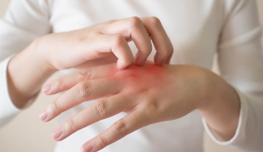
Gout
Gout is a common and complex form of arthritis that can affect anyone. It is characterized by sudden, severe attacks of pain, swelling, and redness in the joints, often the joint at the base of the big toe.
Symptoms
- Severe joint pain. Gout usually affects the large joint in your big toe, but it can occur in any joint.
- Inflammation and redness.
- Limited range of motion.
Reasons
Gout occurs when urate crystals build up in the joints, causing the inflammation and intense pain associated with gout attacks. Urate crystals form when uric acid levels in the blood are high.
Risk factors
You are more likely to develop gout if you have high levels of uric acid in your body. Factors that increase uric acid levels in your body include:
- Diet: A diet rich in meat increases the risk.
- Obesity: If you’re overweight, your body produces more uric acid.
- Medical conditions: Certain diseases and conditions increase your risk of developing gout. These include untreated high blood pressure and chronic conditions such as diabetes, metabolic syndrome, and heart and kidney disease.
- A family history of gout.
- Age and gender: Men are more likely than women to develop gout, usually between the ages of 30 and 50.
Complications
- Recurrent gout
- Advanced gout
- Kidney stones
Diagnosis
- Joint fluid test (uric acid crystals)
- Blood test for elevated uric acid levels
- X-rays
- Ultrasound
Treatment
Medications to treat gout attacks
- Nonsteroidal anti-inflammatory drugs (NSAIDs)
- Colchicine. Your doctor may recommend colchicine, as well as colchicine (Aloprim, Lopurin, Zyloprim) and febuxostat (Uloric), which reduce the amount of uric acid.
Lifestyle and Home Remedies
Limit alcoholic beverages and drinks sweetened with fruit sugar (fructose).
- Limit your intake of foods rich in purines, such as red meat, offal, seafood, and legumes.
- Exercise regularly and lose excess weight.
knee osteoarthritis (OA)
Also known as degenerative knee disease,
OA is typically the result of the gradual wear and tear of articular cartilage.
It is most common in women and elderly men. Knee osteoarthritis can be divided into two types: primary and secondary.
Primary OA is joint degeneration without any clear underlying cause.
Secondary OA is the result of either abnormal concentration of force across a joint, such as post-traumatic causes, or abnormal articular cartilage, such as rheumatoid arthritis (RA). OA is a progressive disease that can eventually lead to disability.
The severity of clinical symptoms may vary from individual to individual. However, they typically become more severe, more frequent, and more debilitating over time. The rate of progression also varies from individual to individual. Common clinical symptoms include knee pain that is initially progressive and worsens with activity, knee stiffness and swelling, pain after prolonged sitting or rest, and pain that worsens over time.
Treatment for degenerative knee osteoarthritis begins with conservative measures and progresses to surgical treatment options when conservative treatment fails.
While medications can help slow the progression of rheumatoid arthritis and other inflammatory conditions, there are currently no proven disease-modifying agents for treating knee osteoarthritis. This activity highlights the role of a multidisciplinary team in caring for patients with this condition. The patient may require physical therapy sessions with specific knee exercises.
Low Back Pain
Low back pain is a common reason for doctor visits.
In Saudi Arabia, the prevalence of LBP among the general population was reported to be 18.8%. Low back pain (LBP) is a significant public health issue, being prevalent and having a significant negative social, psychological, and economic impact.
: Low back pain is a common disorder affecting the muscles and bones of the back. Patients ranged in age from 15 to 52 years. The condition can be further classified by its underlying etiology as mechanical, non-mechanical, or referred pain.
According to the National Institute of Neurological Disorders and Stroke (NINDS), low back pain is the most common cause of job-related disability. At least 80% of Americans experience low back pain in their lifetime.
Spondylosis is a degenerative disorder of low back pain
- Disc injury
- Sciatica
- Spinal stenosis
- Abnormal spinal curvatures
- There are a number of other conditions that cause lower back pain. These include:
- Arthritis is inflammation of the joints
- Fibromyalgia is long-term pain and soreness in the joints, muscles, and tendons
- Spondylitis is inflammation of the joints between the bones of the spine
How is lower back pain diagnosed?
By requesting a complete medical history and performing a thorough physical examination to determine where you’re experiencing pain. The physical examination can also determine if the pain is affecting your range of motion.
Imaging tests such as X-rays, CT scans, ultrasounds, and MRIs may be necessary so your doctor can check for the following:
- Bone problems
- Disc problems
- Problems with the ligaments and tendons in your back
Electromyography (EMG) or nerve conduction tests can help identify any problems with your nerves.
Lower back pain treatment
- Medications
- Over-the-counter (OTC) pain relievers. Nonsteroidal anti-inflammatory drugs.
- Muscle relaxants.
- Topical pain relievers.
- Drugs.
- Antidepressants.
- Physical therapy.
- A physical therapist can teach you exercises to increase your flexibility.
Plantar Fasciitis
Plantar fasciitis is the most common cause of heel pain, according to the American Academy of Orthopaedic Surgeons.
The condition occurs when the plantar fascia at the bottom of the foot becomes inflamed. This ligament is responsible for supporting the arch of the foot.
Causes
Plantar fasciitis is an inflammatory condition that usually has no clear cause. Doctors refer to this condition as idiopathic plantar fasciitis.
Risk factors include obesity, having a very high arch, tight calf muscles, and participating in activities that repeatedly put pressure on the heel, such as running.
Symptoms
Plantar fasciitis causes pain at the base of the heel. This pain is usually worse first thing in the morning when getting out of bed. The pain also worsens with activity.
Treatment
Most people can manage plantar fasciitis with home treatment. Resting the foot and applying ice can reduce inflammation. Nonsteroidal anti-inflammatory drugs (NSAIDs), such as ibuprofen or naproxen sodium, can help manage pain.
Properly stretching the foot before and after physical activity, as well as throughout the day, can help reduce heel pain. Wearing supportive shoes can also help.
If an individual’s plantar fasciitis doesn’t improve with these treatments, they can try physical therapy or visit a podiatrist or orthopedic surgeon for further treatment.
Steroid injections may be recommended to reduce inflammation, or a doctor may prescribe custom orthotics to provide additional heel support.
Osteoporosis
Osteoporosis causes bones to become weak and brittle—making them so fragile that even a fall or minor stresses like bending or coughing can cause a fracture. Fractures associated with osteoporosis most commonly occur in the hip, wrist, and spine.
Bone is living tissue that is constantly being broken down and replaced. Osteoporosis occurs when the creation of new bone does not keep pace with the loss of old bone.
Symptoms
- Back pain due to vertebral fractures or erosion
- Shortness over time
- Easier bone fractures than expected
Causes
Your bones are constantly being renewed—new bone is being made and old bone is being broken down. When you were young, your body made new bone faster than it broke down old bone, thus increasing bone mass. After your early twenties, this process slows down, and most people reach peak bone mass by their thirties. As you age, bone mass is lost faster than it is built.
Immutable risks
- Your gender. Women are more likely to develop osteoporosis than men.
- Age. The older you are, the greater your risk of developing osteoporosis.
- Race. You have a higher risk of developing osteoporosis if you are white or of Asian descent.
- Family history. Having a parent, brother, or sister with osteoporosis puts you at greater risk.
- Body size. Men and women with small body sizes are at greater risk because their bone mass typically decreases with age.
Nutritional factors
- Low calcium intake. Lifelong low calcium intake plays a role in the development of osteoporosis. Calcium intake contributes to reduced bone density, premature bone loss, and an increased risk of fractures.
- Eating disorders. Severely restricting food intake and remaining underweight weaken bones in both men and women.
Medical conditions
The risk of developing osteoporosis is higher in people with certain medical conditions, including:
- Celiac disease
- Inflammatory bowel disease
- Kidney or liver disease
- Cancer
- Lupus
- Rheumatoid arthritis
Complications
Bone fractures, particularly in the spine or hip, are the most serious complication of osteoporosis. Hip fractures are usually caused by a fall and can lead to disability and even an increased risk of death within the first year after the injury.
In some cases, spinal fractures can occur even if you haven’t suffered a fall. The bones that make up the spine (vertebrae) can weaken to the point of shrinkage, which can lead to back pain, decreased height, and a hunched posture in the spine.
Prevention
Good nutrition and regular exercise are essential for maintaining healthy bones throughout your life.
- Protein
- Body weight
Calcium, Vitamin D
Exercise.
Tendonitis and its types: –
- De Quervain’s tenosynovitis
- Trigger finger
- Stenosing tenosynovitis
De Quervain’s tenosynovitis is a painful condition that affects the tendons on the thumb side of your wrist. If you have De Quervain’s tenosynovitis, it will likely hurt when you turn your wrist, grasp something, or make a fist.
- Pain near the base of the thumb
- Swelling near the base of the thumb
- Difficulty moving the thumb and wrist when doing anything that involves clenching or pinching the fingers
- A “tingling” or “cracking” sensation in the thumb when moving it
Treatment: –
The first stage of treatment is to use anti-inflammatory drugs with a pain reliever and a thumb brace. If there is no response to treatment, steroids are injected into the tendon. If there is no response to tendon injections, surgical intervention is performed.
Trigger finger:
It’s a condition in which one of your fingers gets stuck in a bent position. Your finger may bend or straighten, making a clicking sound—like pulling a trigger.
Trigger finger is also known as “trigger finger.” This condition occurs when inflammation narrows the space within the sheath that surrounds the tendon in the affected finger. If trigger finger is chronic, your finger may become stuck in a bent position.
People whose jobs or hobbies involve repetitive actions that require gripping are at greater risk of developing trigger finger. This condition is also more common among women and people with diabetes. Treatment for trigger finger varies depending on the severity of the condition.
Symptoms
- Finger stiffness, especially in the morning
- A popping or crackling sensation when moving the finger
- Pain or a lump (nodule) in the palm of the hand at the base of the affected finger
- Finger stuck in a bent position with a popping sound when suddenly straightened
- Finger stuck in a bent position with an inability to straighten it
Treatment
Steroid injections. Injecting steroid medication near or into the tendon sheath may reduce inflammation and allow the tendons to glide freely again. This is the most common treatment and is usually effective for a year or more in most people treated. However, sometimes more than one injection is required, along with the use of a thumb brace.
Percutaneous release. After numbing the palm, your doctor inserts a strong needle into the tissue around the affected tendon. Moving the needle and your finger helps loosen the blockage that’s impeding the smooth movement of the tendon.
lateral epicondylitis
Tennis elbow, also known as tennis elbow, is characterized by pain on the outside (lateral) side of the elbow. The pain results from damage to the tendons that bend the wrist backward away from the palm. A tendon is a tough cord of tissue that connects muscles to bones. The tendon most likely involved in tennis elbow is called the extensor carpi radialis, and this condition is usually diagnosed in both men and women between the ages of 30 and 50.
Treatment for tennis elbow involves stopping the activity that produces the symptoms. It is important to avoid the movement that caused the injury in the first place. Treatment may include:
- Ice pack application (to reduce inflammation)
- Strengthening exercises
- Anti-inflammatory medications
- Corticosteroid injections
- Surgery